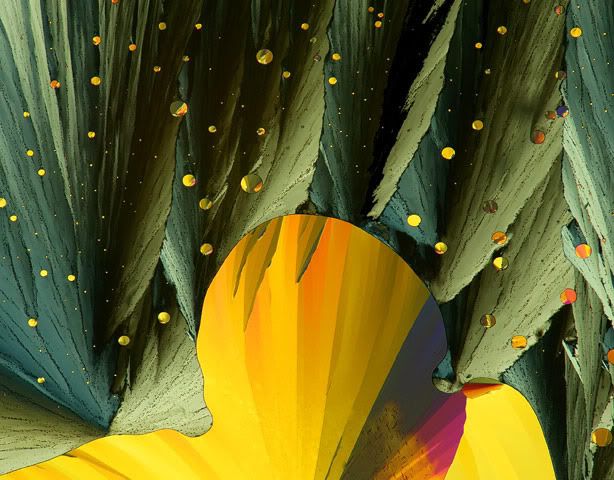
It's that time of the year again! Submissions are in and the 2011 winners are due to be announced soon. Amongst many photo competitions this is my favorite. While I wait for the judges to browse through the candidates I wanted to share few of last year's winners.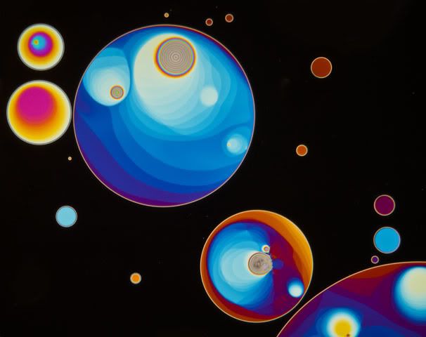
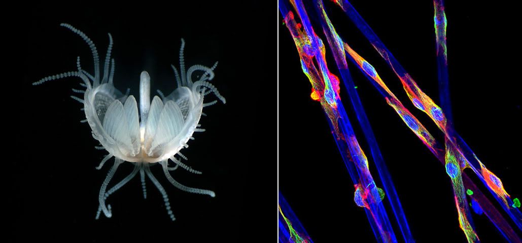
7th Place (right): Endothelial cell attached to synthetic microfibers, stained with microtubules, F-actin and nuclei (2500x). Fluorescent confocal microscopy by Yongli Shan (The University of Texas Southwestern Medical Center, Dallas, Texas, USA).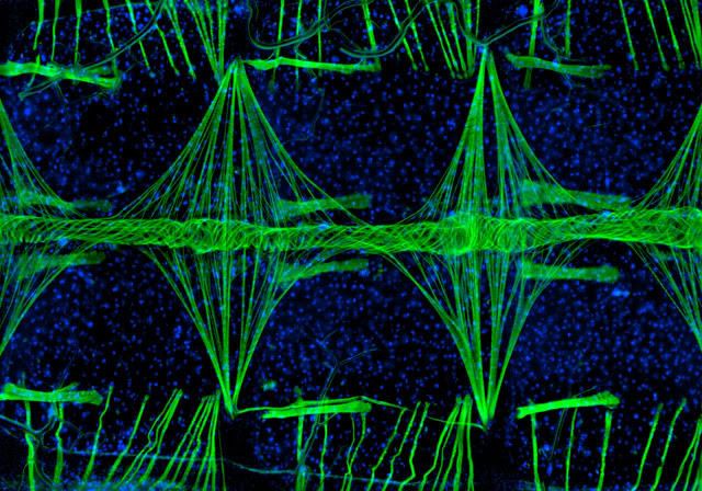
Microscope Photography II
Monday, May 30, 2011 | Posted by Issac Rhim at 8:01 AM | Labels: photography, technology
Subscribe to:
Post Comments (Atom)

2 comments:
have you submitted any?~
no, i wish. haha. i work at a dry lab - no cells to probe around nor a microscope hanging around to look into. =P
Post a Comment